


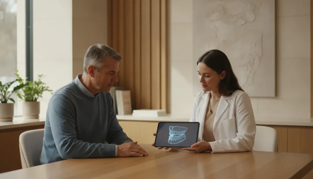
Dental bone graft healing stages describe how a graft turns from a blood clot into bone that can hold an implant. The gum can look closed in a few weeks. The bone underneath is still changing for months. A date on a calendar does not tell you the graft is ready.
Schedule a consultation with Dr. Jefferson Clark, a board-certified prosthodontist in Roseville, to review imaging and plan the next step.
What dental bone graft healing stages actually describe
A dental bone graft is placed where the jaw does not have enough height or width for the planned tooth. The graft is a scaffold. Your own cells have to grow into it, build mineral, and then remodel that mineral into bone that can take a load.
Published reviews of guided bone regeneration describe a sequence, not a single event. In the first day the space fills with a blood clot that releases signals for healing cells. Granulation tissue and new blood vessels replace that clot. Those vessels carry cells that lay down osteoid, the soft framework of bone. Mineral then turns osteoid into woven bone, and woven bone is later replaced by stronger lamellar bone. A 2014 review in The Open Dentistry Journal places that maturation about 3 to 4 months after surgery. Defect size and blood supply change the pace (Liu and Kerns, 2014).
That sequence is the useful meaning of dental bone graft healing stages. Early comfort, a closed gum line, and a graft that is ready for an implant are three different milestones. Mixing them up is why two people with the same surgery date can be on very different plans.
For a week-by-week recovery diary, including swelling, diet, and what the site feels like, read the dental implant bone graft healing process. This page stays with the biology and with what changes the clock.
How the dental bone graft healing stages progress
Think of four overlapping stages. They do not start and stop on a fixed day. A biopsy study of human extraction sockets found wide variation in when mineralized bone appears (Trombelli et al., 2008).
Stage 1: Clot and early inflammation
Right after surgery the grafted space is filled by a clot. Platelets release growth signals, and neutrophils and macrophages arrive to clear debris. This is normal healing, not a sign that the graft has failed. Mild swelling and a dull ache are common in these first days. A sudden increase in swelling, fever, or a bad taste is a reason to call the office, not a stage to wait out.
Stage 2: Provisional matrix and new vessels
The clot is replaced by a provisional matrix, a soft tissue rich in new blood vessels. Human socket biopsies taken at 2 to 4 weeks still showed a large share of granulation tissue. By 6 to 8 weeks that tissue had given way to provisional matrix and woven bone, and osteoblast activity peaked in that window. The same study found that the timing of mineralized bone was much less predictable than the early soft-tissue fill. A site that looks quiet on the surface can still be building this matrix.
Stage 3: Woven bone
Woven bone is the first mineralized bone in the graft. It is real bone, but it is loosely organized and not the tissue you want carrying a crown yet. It acts as a template. New bone is laid onto it in thin layers. On a healing extraction socket, much of this early fill is underway within the first couple of months, while the outer shape of the ridge is still changing.
Stage 4: Remodeling into load-bearing bone
Remodeling replaces woven bone with lamellar bone and marrow. This is the stage that decides implant timing. A European consensus on extraction sites separates two ridges. One has had time for most of this change, often around 12 to 16 weeks. A fully healed ridge is left longer than 16 weeks (Tonetti et al., 2019). Those ranges are for extraction sites managed in a planned way. A larger ridge build, or a site with thin covering gum, can need longer. Dr. Clark confirms readiness with imaging and a clinical exam, not with a date alone.
| Stage | What is happening | What you may notice | What it does not mean |
|---|---|---|---|
| Clot and inflammation. | Clot fills the space and healing cells arrive. | Swelling and a dull ache for a few days. | The graft has already become bone. |
| Provisional matrix. | New vessels replace the clot. | The gum looks quieter. | An implant can be placed yet. |
| Woven bone. | First mineralized bone forms. | The site often feels settled. | The bone is ready for a final crown. |
| Remodeling. | Woven bone becomes lamellar bone. | Little day-to-day change. | Every graft shares one finish date. |
Why two grafts on the same calendar heal differently
The stage names stay the same. The time spent in each stage does not. Four factors move the clock more than willpower or a strict soft-food diet.
Defect size and walls. A socket with most of its bony walls still standing is an easier healing space than a flat ridge that has to be rebuilt in height and width. Fewer walls means less nearby bone to supply cells and blood vessels. Guided bone regeneration uses a membrane to keep soft tissue out while that slower fill happens. Reviews of the method list four conditions: a stable clot, space under the membrane, primary closure of the gum, and exclusion of faster-growing soft tissue.
Graft source. Bone taken from your own body brings living cells, but it requires a second surgical site. Processed human donor bone and animal-derived or synthetic substitutes act mainly as a scaffold. Your cells still have to grow in. A scaffold-only graft can need a longer remodeling window than a small graft that already contains living bone. Neither type comes with a guaranteed finish month.
Blood supply. After a tooth is removed, the ligament that used to feed the socket is gone. The ridge then depends on vessels from the gum and from the bone itself. Surgeons sometimes make small holes in dense cortical bone so marrow blood can reach the graft. A thin flap, heavy smoking, or uncontrolled diabetes can limit that supply. The office will talk through those risks before surgery rather than promise a standard recovery.
Whether the implant goes in now or later. A small defect around an implant that is already stable can sometimes be grafted at the same visit. A site that cannot hold the implant in the right position is grafted first, then the implant is placed after the graft has remodeled. Choosing the staged path is a planning decision. It is not a sign that healing has gone badly.
Where these stages sit in the implant plan
Healing stages answer a planning question: when is this site ready for the next prosthetic step? They do not replace the exam that decides whether you need a graft at all. Imaging of height, width, and the planned crown position comes first. The bone graft for dental implants guide covers that decision, the materials, and what the procedure visit is like.
Timing research sorts implant placement after an extraction into windows. Immediate placement is within about 10 days. Early placement waits about 4 to 8 weeks for soft tissue to close. Delayed placement is often 12 to 16 weeks, after much of the ridge change has occurred. A conventional healed site is left longer than 16 weeks. Ridge preservation, placing a graft at extraction to limit shrinkage, is a modification of the later windows. It does not skip remodeling.
If the tooth is still in place, the extraction visit and the graft are often planned together. The tooth extraction and bone graft overview explains that combined visit. If the tooth has already been gone for years, the ridge may need a larger build, and the remodeling stage is usually the long part of the plan.
Readiness for the implant is not the same as readiness for the final crown. Even after a graft has turned over, a new implant still needs time to lock to bone, commonly discussed as a 3 to 6 month osseointegration window. The final prosthesis is planned after that, in the in-house lab. The temporary and the final teeth are shaped to the bite, not forced onto a site that is still consolidating.
- Confirm the defect with imaging and the planned tooth position.
- Place the graft, alone or with the implant if the site is stable.
- Protect the clot and the closure while the provisional matrix forms.
- Recheck the site before moving from woven bone toward implant placement.
- Build the temporary and final restoration only after integration.
What you can judge at home, and what needs an exam
You can track the early stage. Swelling that peaks in the first few days and then settles, a site you can keep clean as instructed, and discomfort that fades are the usual path. You cannot see woven bone or remodeling in a mirror. A graft can feel fine and still be weeks away from an implant, or it can look quiet and still need a closer look.
Call the office promptly for swelling that gets worse after it had improved, fever, pus, a graft particle flow that does not stop, or numbness that is new. Those are not stages. They are reasons to examine the site. Avoid smoking, straws, and hard chewing on the graft while the clot is still organizing. Those instructions protect stage 1 and stage 2. They do not speed stage 4.
Follow-up visits are how the plan stays honest. Dr. Clark uses them to check closure, comfort, and, when the calendar says a stage might be finishing, the imaging that shows whether mineralized bone is actually there. The practice is out of network with insurance. Visits are reserved for that exam and for the lab work, not for a rushed single sitting.
Schedule a consultation if you have been given a graft timeline and want a specialist to match it to your own imaging.
Frequently Asked Questions
How long do dental bone graft healing stages take?
There is no single finish line. Early soft-tissue fill is often well underway within 6 to 8 weeks. Maturation toward lamellar bone is often discussed around 3 to 4 months for many grafted sites. Extraction-site protocols often re-enter the ridge around 12 to 16 weeks. Larger builds can take longer. Your imaging sets the date.
Does a healed gum mean the graft has finished?
No. Gum closure is an early milestone. Woven bone and later remodeling happen underneath, after the surface already looks quiet. An implant decision waits on that deeper change, not on how the gum looks in a photo.
Can the implant be placed at the same time as the graft?
Sometimes. A small gap beside an implant that is already stable can be grafted at the same visit. If the ridge cannot hold the implant in the right position, the graft is placed first and the implant waits until remodeling has produced enough bone.
What changes the timeline the most?
Defect size, the graft material, blood supply, and medical factors such as smoking or poorly controlled diabetes move the clock more than small differences in home care. Home care protects the clot. It does not replace an exam when the next surgical step is due.
Who should I see to interpret my stage?
A prosthodontist plans the graft backward from the final teeth, not forward from a generic month count. Dr. Jefferson Clark is a board-certified prosthodontist in Roseville and sees patients from Rocklin, Granite Bay, Folsom, and the Sacramento area. A consultation is the right time to match these stages to your scan.
Plan the next step from your own imaging
Dental bone graft healing stages are a map of clot, matrix, woven bone, and remodeling. They explain why a comfortable site can still be weeks from an implant, and why two grafts do not share one calendar. The way to use the map is to put your scan next to it.
Dr. Clark will review the ridge, the planned teeth, and whether this site is still in an early stage or ready for the next one. Care is private and out of network, with time set aside for that planning and for restorations made in the in-house lab.
Schedule a consultation at the Roseville office to review your graft timing.